Selasa, 24 Mei 2011

Theories about the cause of benign prostatic hypertrophy

| Selasa, 24 Mei 2011 | 2 komentar

Theories about the cause of benign prostatic hypertrophy
There are 3 theories put forward about the causes of benign prostatic hypertrophy, are:
1. Stem cell theory
In normal circumstances periuretral gland balanced between growth and the dead (SteadyState). New cells are usually grown from stem cells, because of some reason like the factor of age, hormonal disorders or other factors then trigger the stem cells proliferate faster so happens hiperplasi periuretral gland.

2. Theory Reawakening
Re-grow tissues such as stem cell growth during embriologik level, so that periuretral network can grow faster than the surrounding tissue.

3. The theory de Hidrotestoteron
This theory says that hiperplasi caused by the change in the balance between testosterone and estrogen. Testosterone produced by Leydig cells located in the two testes by 90% while 10% in the adrenal gland. However, there were 20% in a state of free testosterone that can cause prostate enlargement. Testosterone that is free to enter into the prostate to penetrate the cell membrane into the cytoplasm of prostate cells to form DHT heseplar acid complex which will affect the RNA that resulted in the synthesis of proteins that can cause cell proliferation.

Readmore..

Senin, 23 Mei 2011

Nursing care in patients with benign prostatic hyperplasia

| Senin, 23 Mei 2011 | 3 komentar

Nursing care in patients with benign prostatic hyperplasia


DEFINITIONS
Benign prostatic hyperplasia is an urgent periuretral gland hyperplasia prostate tissue native to the peripheral and the surgical capsule.

ETIOLOGY
The cause of benign prostatic hypertrophy is less clear but there are 3 indications that the hormone causes tissue hyperplasia and stromal buffer glandural elements of the prostate. As we get older there will be changes in the balance of testosterone and estrogen, decreased testosterone production and testosterone is converted to estrogen in peripheral adipose tissue. Lobes that have an enlarged neck can clog vesika or prostatic urethra. Thing that happens is that delays the emptying of urine (urinary retention). The result is dilatation of the ureter (hidroureter) and kidneys (hydronephrosis) in stages. Urinary tract infections can occur due to stasis of urine, because most of urine inside the bladder is settled will be where the growth of the organism.

There are several theories about the balance of hormones that cause benign prostate hiperplasi.
1. stem cell theory (Isaacs)
2. Reawekening Theory (Mc Neal)
3. theory de hydro testosterone (DHT)
 
Predisposing factors :
  • Age
  • Hormonal
  • Precipitation factor
  • Too much sitting
  • Too much sex
  • Many masturbate
  • Stress
PATOFISIOLOGY
Areas often affected are the lateral lobe regions of central and medial lobes. Prostate weight is about 60 -100 grams. Enlarged prostate cause narrowing of the lumen of the prostatic urethra pars and will inhibit the flow of urine, causing pressure intervasikal this situation. To remove the urine, bladder must contract more strongly for resistance against it. The contractions which continue to cause anatomic changes of bladder muscle hypertrophy destrusor form, trabekulasi, trbentuknya selula, sakula and divertikel vesika urinaria. At this stage destrusor muscle thickening called phase compensation.
With the increase in urinary retention, detrusor muscle into the phase of decompensation and eventually no longer mapu to contract. Resulting in urinary retention. The pressure of the higher intra vesikal, will be forwarded to all parts of vesika urinaria up in the two estuaries ureter. The pressure on these two estuaries ureter can cause backflow of urine from the bladder into the ureter or bladder occurs fesiko-ureteric reflux. if this situation continues to cause hidroureter, hydronephrosis can occur even kidney failure.

CLINICAL MANIFESTATIONS
Obstructive and irritating symptoms of the syndrome is also called prostatismus the mark with:
  • Increased urinary frequency
  • Nocturia
  • The urge to urinate constantly
  • Abdomen tense
  • Decreased urine volume and must be straining during micturition
  • Urine flow is not smooth
  • Feeling like the bladder does not empty properly.
  • Dribbling (urine continues to drip after urination)
  • Acute urinary retention
  • Recurrence of urinary tract infection
Clinically the degree of symptoms prostatismus divided into:
1. Grade I: symptoms prostatismus + residual urine <50 ml
2. Grade II: symptoms prostatismus + residual urine> 50ml
3. Grade III: urinary retention with upper urinary tract disorders + residual urine> 150 ml

In benign prostatic hypertrophy symptoms of symptoms known as the lower urinity symtoms tract (LUTS), which are divided into obstructive and irritating symptoms.
1. Irritating symptoms
  • Frequent urination
  • Waking at night to urinate
  • Urgency
  • Dysuria
2. Obstructive symptoms
  • Flow / poor stream when urinating
  • Not satisfied in micturition
  • Long wait (hesitancy)
  • Must straining
  • Piss off break
  • long time urinating and urinary incontinence due to overflow.
On the other references are divided into:
1. Grade I (congestive)
  • Increased urination frequency
  • nocturia
  • Difficulty in starting and ending piss
  • Piss off
  • Dribbling
  • poor stream
  • Pain when urinating
  • Pain has not been satisfied after urination
2. Grade II (Residual)
  • Within a few months or years later the patient difficult urination or straining while urinating
  • Urine is not satisfied
  • Urine drips
  • Nocturia
  • Can not urinate
  • Urinary tract infection due to residual urine in vesika
  • High body temperature and potential death
  • Pain in the kidney and spread to the hips
3. Grade III (Urine Retention)
  • Ischuria paradorsal
  • Incontinential paradorsal
4. Grade IV
  • Full bladder
  • Patients in pain
  • Periodic urine dripping (overflow incontinensia)
  • With this infection the patient may die with high heat up to 40-41 degrees Celsius
  • Decreased consciousness
  • Coma
If the fourth grade is not handled properly, it will eventually happen azotamia (accumulation of nitrogen waste products) and renal failure with urinary retention and a large residual volume.


EXAMINATION SUPPORT
  1. Urinalysis
  2. Urodinamis examination (assessing obstruction of urine flow pattern)
  3. Complete blood examination
  4. Assessment of cardiac and respiratory function
  5. Radiological examinations plain to see an enlarged prostate
  6. Ultrasonography (TRUS-transrectal ultrasonography)
  7. Sistografi
MANAGEMENT
  1. Catheterization (with a metal catheter)
  2. Prostatekmi
  3. Watch ful waiting (prostate incision intrasuretral / TUIP)
  4. Balloon dilatation
  5. Gap alfaInhibitor 5 - @-reductase

ASSESSMENT
1. Circulation
Blood pressure rises as the effects of kidney enlargement
2. Elimination
Decrease in strength or encouragement of urine, difficult start, urination, not satisfied in urination, nocturia, Dysuria, Hematuria, recurrent urinary tract infections, urinary tract stone disease history, Constipation
3. Enter the food / beverage
Anorexia, nausea, vomiting, weight loss.
4. Comfort
Suprapubis pain and back pain / hip
5. Security
Fever
6. Sexuality
Decrease in ejaculation

NURSING DIAGNOSIS
1. acute pain associated with muscle spasm spincter
2. changes in the pattern of elimination: urinary retention associated with secondary obstruction
3. Sexual dysfunction associated with loss of body function
4. Risk of infection associated with port de enter microorganisms through a catheter

Readmore..

Rabu, 11 Mei 2011

Nursing care in patients with diarrhea

| Rabu, 11 Mei 2011 | 3 komentar

DIARRHEA

courtesy by e d u
UNDERSTANDING
Diarrhea is the number of bowel movements with stools more than normal (normal 100-200 ml per hour) with the form of liquid or semi-liquid feces, can also be accompanied by an increased frequency defecation.
According to WHO diarrhea is watery bowel movements and more than 3 times a day.
Acute diarrhea is a sudden diarrhea and short duration, within a few hours to 7 days or until 14 days.
Chronic diarrhea is diarrhea that lasts more than 3 weeks.

ETIOLOGY
The cause is a bacterial diarrhea, parasites, or viruses (E.colli, V. cholerae, Aeromonas.SP.)
Another cause is a toxin, drug, food, chemotherapy, fecal impaction and other conditions.

CLINICAL MANIFESTATIONS
Patients with infections often experience diarrhea nausea, vomiting, abdominal pain to stomach cramps, fever and defecation increases with increasing fluid content in feces. spasmodic contraction of the pain and stretching is not effective in anal (tenesmus) can happen every defecation. lack of fluid causes the patient to feel thirsty, tongue dry, and decreased skin elasticity.
hypovolemic cause rapid pulse, decreased blood pressure, nervous, pale, cyanosis, in certain circumstances cause hypokalemia which which will lead to cardiac arrhythmia.

PATHOPHYSIOLOGY
Infectious diarrhea is usually transmitted through the fecal oral because of contaminated food or beverages feces excretion bad plus, the food is not cooked or undercooked. Transmission is person to person via aerosolization or contaminated hands.
Non-invasive bacteria produce a toxin that bound to the intestinal mucosa and increase levels of cyclic AMP in the cell causes active secretion of chloride anions into the intestinal lumen at follow water, carbonate ions, sodium and potassium cations.
Entero-invasive bacteria cause damage to the intestinal wall in the form of necrosis and ulceration, are secretory eksudatif. The liquid can be mixed with mucus and diarrhea with blood.

EXAMINATION SUPPORT
  • Laboratory tests are complete
  • Examination of blood gas analysis
  • Examination of urine and feces

MANAGEMENT
  • Rehydration as a priority treatment. on acute diarrhea can be given ORS, RL fluid or an isotonic Nacl plus 1 ampoule of 7.5% Sodium bicarbonate 50 ml.
  • Identify causes of diarrhea
  • Symptomatic therapy. anti-intestinal motility and secretion (loperamide / Imodium), anti-emetic (metokloperamide).
  • Definitive therapy. cotrimoxazol, cloramphenicol, metronidazole, Ampisillin.

NURSING MANAGEMENT
  1. Measures to control diarrhea by resting in bed, eating and drinking low-fiber to reduce the acute period, limit drinking caffeine and carbonates which stimulates intestinal motility.
  2. Maintaining fluid balance and encourage oral rehydration such as water, juice and broth.
  3. Reduce anxiety by supporting individual coping.
  4. perianal skin care to maintain cleanliness and humidity.
  5. Prevent infections that may occur and prevent the spread of disease through the hands, clothing, bed sheets and other objects.
  6. Monitoring and management of potential complications with the examination of serum electrolyte levels, vital signs and general condition.

COMPLICATIONS
  • Metabolic acidosis
  • Hypovolemic shock
  • Cardiac arrhythmias
  • Acute renal tubular necrosis

NURSING
  1. Physical examination: Patient's general condition: the objective data: body weakness, nausea, vomiting, abdominal pain. Subjective Data: decreased skin elasticity, cyanosis, pale, cold ektrimitas, anuria.
  2. Abdomen: abdominal cramps, hyper peristalsis.
  3. Skin: cold sweat, moisture, changes in skin elasticity.
  4. Neurological: decreased consciousness, seizures.
  5. Psychological: anxiety.
  6. Cardiovascular: Tachicardi, cardiac arrhythmia, shock.

NURSING DIAGNOSIS
  1. Diarrhea associated with the infection process
  2. Imbalance nutrition: less than the needs of biological factors associated with nausea and vomiting
  3. Fluid deficit associated with loss of fluid volume excess
  4. Anxiety associated with changes in health status
  5. Acute pain related to biological factors increasing intestinal motility

Readmore..

Senin, 09 Mei 2011

Body care in AIDS patients

| Senin, 09 Mei 2011 | 0 komentar

Body care in AIDS patients
courtesy by E D U

A. Actions in treatment room
  • Align the patient's body, close your eyes, ears and mouth.
  • Remove the medical equipment that is still attached.
  • Each wound must be in plaster and covered well.
  • Close all holes of the body with waterproof plasters.
  • Clean bodies carefully.
  • Give family and friends to view the corpse.
  • Enter the corpse into a special plastic bag and attach the label "INFECTIOUS / HIV".
  • After all the neat corpse may be sent to the morgue.

B. Actions in the morgue
  • The body was bathed by a morgue worker who has been trained, then is wrapped with a cloth shroud or other packaging in accordance with the belief / religion, and then inserted into a special plastic bag labeled "infectious / HIV".
  • Bodies that have been in special plastic bags should not be opened.
  • The body should not be preserved in any manner or autopsy.
  • In certain circumstances, an autopsy can only be done after obtaining approval from the head of the hospital.

Readmore..

AIDS Patient Care

| | 0 komentar

AIDS Patient Care
courtesy by E D U

The principles of Treatment:

ISOLATION
Basically, AIDS patients can be treated in the usual space infection. Sometimes AIDS patients need care in one special room for consideration:
  • Protect AIDS patients from other infections both endogenous and exogenous.
  • Facilitate the provision of nursing care.

REGULATION OF OFFICERS
a. Nursing team who suffered from infections, skin disorders and pregnant women should not be treating AIDS.
b. Wearing Barakshort (long underwear), masks and eye coverings and gloves for taking action:
  • Bathing.
  • Helping defecation and urination.
  • Preparing laboratory materials.
  • Changing clothes and loom.
  • Expenditure measure urine, faeces or vomit.
  • Doing huknah / klisma and catheterization.
  • Perform procedures medical / invasive: infusion set, clearing the airway, injection and others.
  • Caring for the body.
c. Wash hands before and after treatment measures despite wearing gloves and using a disinfectant under running water.
d. Reporting to the special team handling AIDS in the hospital when exposed to needle puncture scars in use in people with AIDS or splashing liquids when exposed to AIDS sufferers in the eyes, mouth or injured body part.
e. Giving plastic coating on the pillow and mattress as a shield so easy to wash.
f. Replacing the label reads "MATERIALS TRANSMITTED / HIV" in all specimen bottles that will be sent to the laboratory.
g. Cleaning the bathroom and treatment room regularly.
h. uphold the official secret.

PREVENTION OF CONTAMINATION AND CLEANING EQUIPMENT
a. All equipment used by people with AIDS should be set apart.
b. Use disposable tools in the tools such as syringes, infusion set, catheter, endotracheal(ET) tube and others.
c. Loom such as mattresses and pillows were given a plastic coating before it is used to prevent the attachment of blood or body fluids. The loom is already in use fed into a special plastic bag, labeled "INFECTIOUS MATERIAL / HIV", then tied and sent to the place of washing.
d. Gloves are used every action, straightening the bed and holding a tool that has been contaminated.
e. Tool is not the kind of disposable, should be sterilized immediately after use.
f. Masks, oxygen hose, and other breathing aids must be sterilized after use.
g. Instruments wound care or medical equipment, once used soaked with disinfectant solution and then sterilized according to the rules of sterilization.

CONTAMINATED MATERIALS
a. All remaining tissue, blood, body fluids, bandages, needles and abbocath(venous catheter) used during the action placed in hard plastic bag, double-layered, puncture resistant, waterproof and special color, and then given the label "MATERIALS TRANSMITTED / HIV" and then burned.
b. Give the guards with plastic alt on the operating table, operating coat, the base of the hands or head if done action intubation / cannulation / extubasi trachea.

PATIENT RIGHTS
a. patients may visit the facilities available in hospitals such as the television room, canteen and others.
b. Patients can receive guests at the time of visit and may be accompanied during the treatment, except in conditions of infection.
c. Patients with severe diarrhea may only use the special toilet.

COUNSELING
a. Counseling is an important aspect that aims to:
  • Provides information on all matters relating to HIV infection which include, among other causes, clinical symptoms, patterns of transmission, prevention and others.
  • Providing psychological and social encouragement to patients and families in dealing with problems of physical and psychological, so the patient can overcome the problems independently.
  • Providing encouragement to the patient to adjust to new conditions, among others by holding a sexual behavior change, to reduce the spread of the HIV virus to others.
b. Counseling done by doctors and or nurses who have been specially trained.
c. Counseling target not only patients and families but all health workers including nurses, physicians, laboratory workers, laundry officers and others.

MAINTENANCE OFFICER ATTITUDES
a. Understand, understand and have skills in providing nursing care.
b. Be calm, fair, and not excessive but remain vigilant in helping patients.
c. Understanding the state of the patient, show empathy.
d. Be protected.

Readmore..

Kamis, 21 April 2011

Nursing care in patients with AIDS

| Kamis, 21 April 2011 | 0 komentar

Courtesy BY E D U
A I D S
UNDERSTANDING
  • HIV (human immunodeficiency virus): The virus that causes one's immune system weaknesses.
  • AIDS (Acquired immunodeficiency syndrome): an infection that can cause severe damage to the immune system and can not be cured, so people are very open or vulnerable to infection or certain cancers.
  • Patients with HIV: is someone who is infected with human immunodeficiency virus, which infects mainly T lymphocyte cells and disrupt cell immunity.
  • Patients with HIV-AIDS is someone who has been infected with HIV and are at the last stage of infection with signs of the emergence of various bacterial infectious diseases, fungi, parasites and viruses that are opportunistic or malignancy.
CAUSE
AIDS is caused by human immunodeficiency virus. This type of retrovirus that is easy to die outside the body. The period between infected with HIV until the onset of symptoms (incubation period) is 6 months in children and 60 months in adults.

SYMPTOMS AND SIGNS
Clinical stage I:
  • Without symptoms (asymptomatic)
  • Generalized lymphadenopathy which persistent
Clinical Stage II:
  • Weight loss <10% - Preview minor mucocutaneous - herpes zoster - Upper respiratory tract infections recurring Clinical Stage III: - weight loss> 10%
  • Prolonged fever that can not be explained (intermittent or constant), more than a month
  • Oral candidiasis
  • Pulmonary Tuberculosis
  • Severe bacterial infection
Clinical Stage IV:
  • HIV wasting syndrome
  • Pneumonia carini
  • Toxoplasmosis of brain
  • CMW in organs other than liver, spleen, or lymph nodes
  • Infection with Herpes simplex virus (HSV) mucocutaneous more than 1 month
  • Candidiasis esophagus, trachea, bronchus, or lung
  • Micobakteriosis
  • Septicemia
  • Extra pulmonary tuberculosis
  • Lymphoma
  • Kaposi's sarcoma
  • HIV encephalopathy
LABORATORY EXAMINATION
1. Tests to determine the presence of antibodies and HIV antigen:
  • Detection of HIV antigen by ELISA (enzyme linked immunosorbent assay).
  • Immunoblot western blot (WB)
2. The tests showed a decrease in body immunity
Decrease the body's immunity:
Simple laboratory tests: Hb, Smear of peripheral blood, leucocytes, lymphocytes and platelets, the AIDS found existence of anemia ,leukopenia ,and thrombocytopenia limfopenia .

3. Testing for the presence of malignancy or opportunistic infection
  • Serology or culture against herpes simplex, CMV, Epstein Barr
  • Staining culture or histopathology of pneumonia carinii, cryptosporidium, toxoplasmosis, candidiasis, or aspergilus.
  • Histopathologic of Kaposi's sarcoma or lymphoma.
NURSING ASSESSMENT IN AIDS PATIENTS
A. Diagnose
History of free sex, homosexuality, history of drug abuse via intravenous blood transfusion recipient, a parent suffering from HIV - AIDS, chronic diarrhea, decreased appetite, weight loss, fever.
B. Psychological
knowledge and acceptance of disease, function and role in family, community, and employment.
acceptance of the patient's family.
C. Social
Support systems are available and affordable with the ability financially.
The Group of HIV-AIDS sufferers.
D. Clinical manifestations
clinical manifestations that spread throughout the body that can of whole organs, infections, malignancies.
E. Neurologic (malignancy)
HIV encephalopathy, Cryptococcus neofarmans, progressive multifocal lauciensefalopaty (PML).

NURSING DIAGNOSIS
  1. PK: Opportunistic Infections
  2. PK: Sepsis
  3. PK: malignancies
  4. Ineffective airway clearance
  5. Diarrhea
  6. Changes in family process
  7. Risk of infection
  8. The risk of transmission of infection
  9. Fatigue
  10. Social isolation
  11. The risk of ineffective management of therapeutic regimen
  12. Powerlessness
  13. Anxiety

Readmore..

Jumat, 08 April 2011

Nursing care in patients with Lung Cancer

| Jumat, 08 April 2011 | 3 komentar

LUNG TUMOR (CARCINOMABRONCHOGENIC)

DEFINITION
Bronchogenic carcinoma is a malignant tumor originating from primary lung airways.
Most of the primary lung tumor is a carcinoma bronchi (John E. Stark, 1990).

PHYSICAL SYMTOMS
Hemopthisis, Cough, Chest pain, Shortness of breath, this is due to enlargement of the tumor and due to collapse of the lung, Wheezing / stridor, this sounds arising from the trachea or bronchus obstruction, Hoarse, this happens due to his stricken recurents left laryngeal nerve, Pneumonia Recurents, Dysfagia, this may occur due to the spread of tumors via lymphatic vessels to the area mediatinum or to the esophagus, Obstruction of superior vena cava, Systemic symptoms: such as weight loss, no appetite, which is the initial symptom in 50% of patients with lung cancer, Symptoms of metastasis, the most common of the organs of the brain, liver, bone and adrenal gland, Non-metastatic effects: such as peripheral neuropathy, dermatomiositis or syndrome whose symptoms such as secretion of hormones (eg ADH, ACTH, PTH).

ETIOPATOGENSIS
Such as cancer in general, the exact etiology of carcinoma bronkogenik still unknown, but it is expected that the long-term inhalation of carcinogenic materials is a major factor, without prejudice to possible predisposing role of family relations or ethnic / racial and immunologis status. Inhalation of carcinogenic materials is highlighted that many cigarettes.


HIGH RISK GROUPS:
- Smokers.
- Workers at asbestos factories.
- History fibrosis suffer from chronic lung diffus.

EFFECT OF CIGARETTE:
The materials are carcinogens in tobacco smoke include: polomium 210 and 3.4 benzypyrene. The use of a filter is said to reduce her risk of carcinoma broncogenik, but still remained higher than non-smokers.
In the long term ie, 10-20 years, smoking:
1-10 cigarettes / day increases the risk 15 times
20-30 cigarettes / day increases the risk 40-50 times
40-50 cigarettes / day increases the risk 70-80 times.

Industry Influence
The most widely associated with the carcinogen is asbestos, which otherwise increases the risk of cancer is 60-10 times. Following the industrial radioactive materials, miners uramium 4 times the population at risk in general. Exposure to this industry only visible effect after the 15-20 years.

Effect of Other Diseases
Pulmonary tuberculosis is associated as much bronchogenik carcinoma predisposing factors, through mechanisms hyperplasi - metaplasi - carcinoma in situ-carcinoma - bronkogenik as a result of scar tissue tuberculosis.

Effect of Genetic and immunological status
In 1954, Tokuhotu can prove that despite the influence of heredity rather than factors of environmental exposure, this provides an opinion that can be derived bronkogenik carcinoma. Research recently skewed that the factors involved with Aryl Hydrocarbon hydroxylase enzymes (AHH). Status immonologis patients are monitored from cellular mediated showed a correlation between the degree of cell differentiation, stage of disease, response to treatment and prognosis.

Classification by histopathology using ordinary light microscope (WHO, 1977).
1. Epidermois carcinoma (squamous cell carcinoma).
2. Adeno carcinoma
3. Undiferentiated small cell carcinoma (oat cell)
4. Large cell carcinoma undeferentiated.

INVESTIGATIONS
Radiological
Radiopaque mass in the lung, Airway obstruction with resultant atelectasis, Pneumonia, Enlarged hilar glands, Cavitation.

Sputum cytology:
In sputum cytologic examination to help establish the case up to 70%. Sputum for cytologic sample should be received by the laboratory within 2 hours after ekspectorasi / expenditure. Sample dawn is not required.

Bronchoscopy:
In the biopsy is used to determine the type of tumor cells. Bronkografi
The picture is considered bronkografi patognomonik irregular stenosis is obstruction, stenosis rats and indented thumb.

Pleural aspiration and biopsy:
Aspiration is an action that must be done if patients with lung tumors have effusi pleura. Effusi not always result from the spread of tumors to the pleura, but may result from pneumonia reaction to the tumor or lymphatic obstruction.

Biopsy needle percutan:
This examination is useful for diagnosing tumors that are difficult peripheral transbronchial biopsied denag techniques.

Mediatinoscopy:
This technique is used to take samples of lymph gland enlargement mediatinum experiencing, this is done if no visible pulmonary tumor.

Endoscopy
Includes examining laryngoscopy and bronchoscopy and bronchial washings, scrapings / sweep and biopsy. The objective examination of Bronchoscopy (fiber optics) are:
a. Knowing the changes in the bronchus of lung cancer.
b. Retrieving material for cytological examination.
c. Noting the changes on the surface of tumor / mucosa to predict the type of malignancy.
d. Assessing the success of therapy.
e. Determining overbilitas lung cancer.

Immunology
The existence of a negative correlation between cancer and immunological reactions have been generally known. Immunological disorders mainly seen in cell mediated immunity that can be given through a delayed hypersensitivity reaction is clearly, tolerance to skin graft, total circulatory low T cell, and lymphocyte transformation in vitro is low. At this time more immunological examination serve as prognostic factors than diagnostic factor. Conclusion Correlation of skin test and response to cytostatic:
a. Less than 1.0 cm. : Prognosis is poor, widespread disease.
b. Less than 2.5 m. ; Better prognosis, limited disease, good response to chemotherapy.

PHASING CLASSIFICATION CLINIC (Clinical Staging)
Based on TNM
T = Tumor: N. : Nodules, namely the lymph nodes of M. : Metastases
1. T: T-0: No visible primary tumor
  • T-1: tumor diameter of less than 3 cm. Without the invasion of bronchus
  • T-2: tumor diameter more than 3 cm. Can be accompanied by atelectasis or pneumonitis, but is more than 2 cm. From Karina, and there is no pleural effusion.
  • T-3: Tumor size with an invasion into the surrounding (thoracic wall, diaphragm or mediastinum) or have been near Karina accompanied by pleural effusion.
2. N: N-0: There was no propagation to regional lymph nodes.
  • N-1: There is a propagation to the ip silateral hilar lymph nodes.
  • N-2: There is a spreading to the lymph limfemediastinum or contralateral
  • N-3: There extratoracal spreading to lymph nodes.
3. M. M-0: There is no distant metastases.
M-1: Already there are distant metastasis to other organs.
Based on TNM. Compiled phasing following clinics.
a. Carcinoma in situ: T-0, N-0, M-0, but positive sputum cytology for malignant cells.
b. Phase I. T-1, N-0, M-0, or T-2, N-0, M-0
c. Phase II. T-2, N-1, M-0.
d. Stage III: when there are already T-3, N-2, or M-1.


MANAGEMENT
Treatment of lung tumors depend on tumor cell types.
1. Surgical resection.
2. Palliative therapy.

ASSASSMENT:
Activity / rest : Weakness, inability, to maintain regular habits, dyspnea because the activity, lethargy usually advanced stage.

Cardiovaskuler and circulation :
Pallor, cyanosis, diaphoresis, hypotension, bradycardi, tachycardi, arrytmia in atrial or ventricular, decreased cardiac output, shock. Increased jugular vein, heart sound: friction pericardial (addressing effusion) Dysrhythmias, finger percussion.

Ego Integrity : Anxiety, fear of death, resist harsh conditions, anxiety, insomnia, the question is repeated. lack of rest.

Elimination :
Diarrhea that intermittent (hormonal imbalance) Increased frequency / amount of urine (Hormonal Imbalance).

Food / liquids :
Weight loss, poor appetite, decreased food input, difficulty swallowing, thirst / increase fluid intake, Thin, wiry, less weight or appearance (stage 0, edema face, periorbital (hormonal imbalance), Glucose in the urine.

Discomfort / pain :
Chest pain, which does not / can be affected by the change of position. Painful shoulder / hand, bone pain / joint, cartilage erosion secondary to the increase of growth hormone. Abdominal pain is gone / arise.

Respiratory :
Cough mild cough or a change from the usual pattern, increased sputum production, shortness of breath, workers exposed to carcinogenic substances, hoarse, vocal cord paralysis, and smoking history. dyspnea, increased employment, increased tactile fremitus, wheezing on inspiration or expiration (air flow interruption). persistent wheezing tracheal deviation (the area that suffered lesions) hemoptysis.
Blood gas analysis (obtained hypoksemia, acidosis, an increase or decrease in CO2). Respiratory function (VC reduction, increased tidal volume). ECG (may show a arrytmia).

Security : Fever, maybe there is / are not, reddish, pale skin.

Sexuality :
Gynecomastia, amenorrhea, or impotence.
family risk factors: a history of lung cancer, tuberculosis.

NURSING DIAGNOSIS
  • Ineffective breathing pattern related to decreased lung expansion.
  • Ineffective airway clearance related to airway obstruction.
  • Damage to gas exchange associated with chronic hypoxia in lung tissue.
  • Anxiety associated with an inability to breathe.
  • Acute pain b / d of cancer invasion into the pleura, chest wall.
  • Nutrition less than body requirements b / Inadequate nutrition, increased metabolism, the process of malignancy.
  • Impaired body image b / d of changes in body structure.

Readmore..

Senin, 04 April 2011

Nursing care in patients with Congestive Heart Failure

| Senin, 04 April 2011 | 1 komentar

Inception Report
NURSING IN PATIENTS WITH CONGESTIVE HEART FAILURE
courtesy edu

UNDERSTANDING
Congestive heart failure is a condition where the heart can no longer pump enough blood to meet body needs circulation for tissue metabolism in certain circumstances, whereas in cardiac filling pressure is still high.

CAUSES OF HEART FAILURE
  1. Causes of heart failure are classified as follows:
  2. Myocardial dysfunction (myocardial failure).
  3. Expenses excessive pressure - systolic loading (systolic overload).
  4. Excessive volume load - loading diastolic (diastolic overload).
  5. Impaired filling (input resistance).
  6. Increased metabolic needs - increasing need for excess
LEFT HEART FAILURE SYMPTOMS:
Complaints of body feeling weak, tired, palpitations, shortness of breath, cough, anorexia, and sweating cold, cough and / or coughing up blood, decreased kidney function.

RIGHT HEART FAILURE SYMPTOMS:
Edema, anorexia, nausea, ascites, abdominal pain.

FUSE
Hypertension, myocardial, pulmonary embolism, infection, arrhythmia, anemia, febrile, emotional stress, pregnancy / delivery, infusion / transfusion.

PATHOPHYSIOLOGY
Each obstacle in the flow direction (forward flow) in the circulation will cause the dam to flow in reverse direction (backward congestion). Barriers flux (forward failure) will cause the symptoms of failure in the system backward flow of blood circulation. Cardiac compensatory mechanisms in heart failure is the way the body to maintain blood circulation to meet tissue metabolic needs. Compensatory mechanisms that occur in heart failure are: ventricular dilation, ventricular hypertrophy, increased sympathetic stimulation in the form of tachycardia and peripheral vasoconstriction, elevation of plasma catecholamine levels, retention of salt and fluid loss and increase oxygen ekstraksi by the network.
When the heart of the right and the left together in a state of failure due to interruption of blood flow and the presence of dams, it would appear the signs and symptoms of heart failure on systemic circulation and pulmonary circulation. This is called Congestive Heart Failure (CHF).

LABORATORY EXAMINATION:
There is no specific test that can diagnose heart failure (T. Santoso, heart failure 1989). Laboratory tests are needed to determine the extent to which heart failure has been disturbing the functions of other organs such as liver, kidney and others.

EXAMINATION OF OTHER SUPPORT:
A. Radiology:
  • The image of the lung hili thick and wide, getting to the edge density decreases.
  • Field lung spots because of pulmonary edema.
  • Pulmonary venous distension.
  • Hidrothorak.
  • Enlargement of the heart, Cardio-thoragic ratio increased.
B. ECG:
Can be found in the primary abnormality of the heart (ischemia, ventricular hypertrophy, rhythm disturbances) and signs of acute trigger factors (myocardial infarction, pulmonary embolism).

C. Echocardiography:
For detection of functional and anatomical disorders that cause heart failure.

D. Cardiac Catheterization:
In left heart failure is obtained (VEDP) 10 mmHg or pulmonary arterial wedge pressure> 12 mmHg in the resting state. Cardiac output is lower than 2.7 lt/mnt/m2 body surface area.

MANAGEMENT
According to the priority is divided into 4 categories:
  1. Improving myocardial contraction / perfusion systemic.
  2. Lowering the volume of excess fluid.
  3. Post Op prevent complications.
  4. Surgical treatment (Komisurotomi).
  5. Health education regarding disease, prognosis, medications and prevention of recurrence.

ad. 1 Improving myocardial contraction / perfusion systemic:
  • Bed rest / bed rest in semi-Fowler position
  • Provide oxygen therapy as needed
  • Provide medical therapy: digitalis to strengthen heart muscle contraction
ad.2 Lose excess fluid volume
  • Provide medical therapy: diuretics to reduce fluid in the tissues
  • Record intake and output
  • Considering weight
  • Restriction of salt / low salt diet
ad.3 Preventing complications
  • Schedule a gradual mobilization according to client circumstances
  • Prevent the occurrence of immobilization due to bed rest
  • Changing sleeping positions
  • Noting the adverse effects of Medica mentosa; digitalis poisoning
  • Checking or monitoring ECG
ad.4 Surgical Treatment Komisurotomi
Only in the aorta due to infection of aortic regurgitation, aortic valve repair can be considered. While in aortic regurgitation due to other diseases should generally be replaced with artificial valves. Indications on the complaint shortness of breath that can not be overcome by treatment symptomatik. When ekhokardiografi showed left ventricular systole 55 mm, or 25% fractional shortning considered for surgery before heart failure arise.

ad.5 health education, in terms of disease, prognosis, drug use and preventing relapse
  • Explaining the course of the disease and prognosis.
  • Explaining the use of medications that are used, as well as providing drug administration schedule.
  • Changing lifestyles / habits that one: smoking, stress, heavy work, drinking alcohol, foods high in fat and cholesterol.
  • Explain about the signs and symptoms that contribute to heart failure, particularly those associated with fatigue, soon tired, palpitations, shortness of breath, anorexia, cold sweat.
  • Advise to all controls on a regular basis even if no symptoms.
  • Providing mental support; client to accept his situation is real / reality will itself well.
ASSESSMENT OF DATA

1. Activity and rest
  • The existence of fatigue / exhaustion, insomnia, lethargy, lack of rest.
  • chest pain, dyspnea at rest or during activity.
2. Circulation
  • History of hypertension, valve disorders, cardiac surgery, endocarditis, anemia, septic shock, swelling in the legs, ascites, tachycardia.
  • Dysrhythmias, atrial fibrillation, premature ventricular contraction.
  • Sound S3 Gallop, the sound of CA, the presence of systolic or diastolic, murmur, increased JVP.
  • Presence of chest pain, cyanosis, pale, ronchi, hepatomegaly.
3. Mental Status
  • Anxiety, fear, anxiety, anger, iritabel / sensitive.
  • Stress related to illness, financial social
4. Elimination
  • Decrease in urine volume, urine is concentrated
  • Nocturia, diarrhea and constipation
5. Food and fluid
  • Loss of appetite, nausea, and vomiting
  • In the lower limb edema, ascites
6. Neurology
  • Dizziness, fainting, pain
  • Lethargi, confusion, disorientation, iritabel
7. Comfort
Chest pain, chronic / acute angina

8. Respiration
  • Dispnoe at the time of activity, takipnoe
  • Sleeping and sitting, history of lung disease
9. Sense of security
  • Changes in mental status
  • Disorders of the skin / dermatitis
10. Social interaction
Reduced social activity

PRIORITY TREATMENT
  • Increase myocardial contractility / systemic tissue perfusion.
  • Lose excess fluid volume.
  • Preventing Post op complications.
  • Provides information about the tailor, prognosis, therapy and prevention of recurrence of disease.
TREATMENT DIAGNOSIS FREQUENTLY ARISING:
  1. Decrease in cardiac output in relation to the decrease in myocardial contractility, characterized by:Increased heart rate, changes in blood pressure, decreased urine output, the S3 and S4, chest pain.
  2. Limitations of activity in relation to the imbalance between supply and demand of oxygen, characterized by: weakness, fatigue, changes in vital signs, dysrhythmias, dispnoe, diaporesis
  3. Disorders of fluid balance, more of the needs in relation to the decline in GFR, marked by: heart sounds 3, orthopnoe, oliguria, edema, weight change, hypertension, respiratory distress, abnormal breath sounds.
  4. High risk of failure of gas exchange in relation to changes in alveolar capillary membrane by accumulation of fluid in the lung cavity.
  5. Risk of damage to skin integrity about its continuity due to edema, decreased perfusion to the skin.
  6. Lack of knowledge about diseases, conditions and treatment in relation to the lack of information is marked with: the patient asked the patient the wrong statement.

Readmore..

Dysphagia

| | 1 komentar

Disfagi
 
Definition
Realize the difficulties in solid or liquid food flowing from the mouth through the esophagus.

Pathogenesis
1. Lumen of the esophagus or oropharynx obstruction due to intrinsic lesions on the wall, extrinsic compression or foreign objects of nature lumen.
Causes include :
  • Malignancy (primary or secondary)
  • Peptic stricture
  • Chemical injury (eg corrosive)
  • "oesophageal web"
  • Esophageal diverticulum
  • Infection of the esophagus (candidiasis)
  • Foreign body (corpus alenum)
  • Vascular (large left atrium)
2. Neuromuscular disorders that interfere with the coordination of the flow of normal food and fluid from the esophagus to the stomach.
causes include:
  • Cerebro vascular accident
  • Motor neuron disease
  • Multiple sclerosis
  • Myasthenia gravis
  • Polimiositis, dermatomiositis, scleroderma
  • Tiro toxic myopathy
  • Akalasia
Clinical picture1. difficulty in cleaning the posterior pharynx, often accompanied by nasal regurgitation and pulmonary aspiration, almost always associated with neuro muscular disorders oropharynx. in such cases, solid and liquid foods can trigger symptoms.

2. Dysphagia for solid and liquid meal in patients who can cleanse the posterior pharynx leading to esophageal disorders such as diffuse esophageal spasm, or sklero akalasia charity. typical dysphagia are intermittent and not progressive.

3. Dysphagia a progressive slow, initially limited to solid foods, in patients with a history of gastro-esophageal reflux before, leading to peptic stricture.

4. Dysphagia rapid progressive, especially in elderly patients, typical for malignant lesions obstruction.

5. Chest pain accompanied by dysphagia have limited diagnostic value and occurs in both the esophageal spasm or obstructive lesions in each.

Diagnosis
Observation of patients swallowing is an important diagnostic maneuvers and should be performed for all patients. When the oropharynx disorder is suspected, formal neurologic examination should be performed with direct visualization of the neuromuscular function of the pharynx and larynx.

Radiology
"Barium swallow" with contrast medium flow visualization carefully is the most important examination for verification for each obstructive lesions. This examination must show proof akalasia and maybe get other neuromuscular disorders, especially spasm of the esophagus.

Endoscopy
Fiber-optic endoscope may be needed to see firsthand and to biopsy lesions that are not entirely typical of the "barium swallow", especially to distinguish between benign and malignant strictures. If endoscopy performed before the "barium swallow" takes meticulous care to avoid trauma and possible perforation of the esophagus with high-lesion location. If the tests above failed to get the diagnosis or lead to motility disorders, esophageal manometri be done.

Readmore..

Rabu, 30 Maret 2011

Nursing care in patients with Acute tonsillitis

| Rabu, 30 Maret 2011 | 4 komentar

courtesy by E D U
Acute tonsillitis
(tonsillectomy) 
Definition
Tonsillitis is the presence of general inflammation and swelling of tonsil tissue with a collection of  leucocytes, el-dead epithelial cells and pathogenic bacteria in kripta (Adam Boeis, 1994: 330).
Tonsillectomy is an invasive measures being undertaken to take the tonsils with or without adenoid (Adam Boeis, 1994: 337).

Etiology
• Hemolitikus streptococcus group A.
• Pneumococcal.
• Staphylococcal.
• Haemofilus influezae.

Pathofisiologi
• The occurrence of inflammation in the area tonsila from the virus.
• Resulted in the formation of exudate.
• Cellulitis occurs tonsila and the surrounding area.
• Peritonsilar abscess formation.
• Tissue necrosis.

Symptoms
• Sore throat and dysphagia.
• Patients do not want to eat or drink.
• Malaise.
• Fever.
• Breath odor.
• Otitis media is one of the originators.

Management
• Bed rest.
• Provision of adequate fluids and light diet.
• Giving medications (analgesics and antibiotics).
• If no progress then the alternative measures that can be done is surgery.

Indications of surgery Indications of absolute
• incidence of pulmonary choir due to chronic airway obstruction.
• Hypertrophy of tonsils or adenoids with apnea syndrome during sleep.
• Excessive hypertrophy resulting in dysphagia and weight loss as a companion.
• Excision biopsy in suspected malignancy (lymphoma).
• Peritonsilaris recurrent abscess or abscess that spread to surrounding tissues.

Relative indication
All other indications for tonsillectomy action is considered as an indication of the relative.
Another indication that most can be received is:
• Recurrent tonsillitis attacks.
• Tonsil hyperplasia with functional disorders (dysphagia).
• Hyperplasia and obstruction which settled for 6 months.
• Not respond to treatment and therapy.

Contraindications
• Fever is not in the know the cause.
• Asthma.
• Systemic infection or chronic.
• Sinusitis.

Preparation of operations that may be done
• Laboratory tests (Hb, leko, bleeding time).
• Give an explanation to the client action and care after surgery.
• Fasting 6-8 hours before surgery.
• Give antibiotics as prophylaxis.
• Give premedication ½ hours prior to surgery. 

Assessment
  • Medical history related to the factor supporting the occurrence of tonsillitis and the bio-psycho-socio-spiritual. Blood circulation : Palpitations, headache at the time of change of position, decreasing blood pressure, bradycardia, body felt cold, pale extremities.
  • Elimination : Changes in the pattern of elimination (incontinence uri / alvi), abdominal distension, bowel sounds disappearance.
  • Activity / rest : There is a decrease in activity because of weakness of body, loss of sensation or parese / plegia, tiredness, difficulty in recuperating from seizures or muscle spasms and pain.The reduced level of consciousness, decreased muscle strength, general body weakness.
  • Nutrition and fluids : Anorexia, nausea, vomiting due to increased ICT (intra-cranial pressure), impaired swallowing, and loss of sensation on the tongue.
  • Nerve supply : Dizziness / syncope, headache, decreased visual field area / blurred vision, decreased sensation of touch, especially in the area face and extremities.
  • Mental status : coma, weakness in the extremities, muscle paralise face, aphasia, pupil dilation, decreased hearing.
  • Comfort : Tense facial expressions, headache, anxiety.
  • Breathing : Shortened breath, inability to breathe, sleep apnea, the occurrence period of apnea in breathing pattern.
  • Security : Fluctuation of temperature in the room.
  • Psikolgis : Denial, disbelief, profound sadness, fear, anxiety.

    Nursing problem and action plan

    Ineffective breathing pattern associated with tissue damage or trauma to the respiratory center
    Objective:
    Patients showed the ability to perform adequately with respiratory blood gas results showed a stable and good and the loss of signs of respiratory distress.

    Plan of action:
    • Exempt a patent airway (keep your head in a state parallel to the spine / as indicated).
    • Perform suctioning if necessary.
    • Assess the respiratory system function.
    • Assess the patient's ability to do cough / business release secretions.
    • Observation of vital signs before and after taking action.
    • Observation for signs of respiratory distress (the skin becomes pale / cyanosis).
    • Collaboration with the therapists in the provision of physiotherapy.

    Impaired sense of comfort pain associated with physical trauma
    Objective:
    Patients express the pain is reduced and shows a state of relaxed and calm.

    Plan of action:
    • Assess the level or degree of pain felt by patients in using the scale.
    • Help the patients in finding the factors the precipitation on pain in feel.
    • Create a peaceful environment.
    • Teach it to a patien and demonstration of several ways of doing relaxation techniques.
    • Collaboration in the provision of appropriate indications

    Readmore..

    Selasa, 29 Maret 2011

    Nursing care in patients with Anemia aplastic

    | Selasa, 29 Maret 2011 | 8 komentar

    Anemia aplastic


    A. UNDERSTANDING
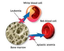
    Aplastic anemia is a disorder in bone disumsum stem cells that can cause death if the amount of blood cells produced are not adequate.

    B. ETIOLOGY
    • Certain antibiotics such as chloramphenicol
    • Viral infections such as hepatitis
    • Benzene
    • Radiation Therapy
    • Antineoplastic agents or cytotoxic
    C. Pathophysiology
    The existence of an anemia reflect the existence of a marrow failure or excessive loss of red blood cells or both. Marrow failure (eg reduced eritropoesis) can occur due to nutritional deficiencies, toxic exposure, invasion of tumor or other cause not yet known. Red blood cells can be lost through bleeding or hemolysis (destruction).

    Red blood cell lysis (dissolution) occurs mainly in phagocytic cells or the reticuloendothelial system primarily in the liver and spleen. As a result of this process is bilirubin that form in the phagocyte will enter the bloodstream. Any increase in red blood cell destruction (hemolysis) immediately reflected by increased plasma bilirubin. (Normal concentration of 1 mg / dl or less; levels above 1.5 mg / dl result in jaundice in the sclera).
    If experiencing the destruction of red blood cells in circulation, the hemoglobin will appear in the plasma (hemoglobinemia). If the plasma concentration exceeds the capacity of plasma haptoglobin (hemoglobin-binding proteins for free) to bind it all (eg if there are more than about 100 mg / dl), hemoglobin will be diffused in the kidney glomerulus and into the urine (hemoglobinuria).
    Conclusions about whether an anemia in certain patients is caused by destruction of red blood cells or red blood cell production is not sufficient in a way that is:
    1. Calculate reticulocyte in blood circulation.
    2. The degree of proliferation of young red blood cells in bone marrow and how pematangannya.
    3. Presence or absence of hyperbilirubinemia and hemoglobinemia.
    Aplastic anemia is caused by a decrease in precursor cells in bone marrow with fat that can occur in congenital or acquired and idiopathic (no cause is not clear). Various kinds of infections during pregnancy can be as originators, or can be caused by drugs, chemicals, or damage radiation. Substances that often cause bone marrow aplasia is benzene and benzene derivatives (eg, airplane glue); anti-tumor drugs such as nitrogen mustard,; antimetabolik, including metotrexate and 6-merkaptopurin; and berabagai other toxic substances such as arsenic.

    Various materials which sometimes causes aplasia or hypoplasia include various antimicrobial, anti-convulsive, anti-thyroid drugs, oral hypoglycemic drugs, anti histamine, analgesic, sedativ, phenothiasine, insecticides, and heavy metals. In various circumstances, aplasia anemia occurs when the drugs or substances included in the amount of toxic chemicals. If exposure is stopped immediately (in this case at the first occurrence of retikulositopenia, anemia, granulositopenia, thrombocytopenia) can be expected soon and perfect healing. In a young man who experienced puberty during hepatitis have a high risk of experiencing severe aplasia anemia with mortality rates of 90% in the first year with a mean survival of six months; marrow transplantation is a treatment option.

    D. CLINICAL MANIFESTATIONS
    • Pale as a result of reduced blood volume and decreased hemoglobin.
    • Dyspnea, shortness of breath and tired quickly when berktivitas.
    • Loss of appetite, nausea and stomatitis.
    • Tachycardia and cardiac noise as a picture of the workload and bulk Increased heart.
    • Headache, dizziness and weakness as a result of reduced supply of oxygen carried by red blood cells into the central nervous system.
    E. EXAMINATION SUPPORT
    Laboratory (Hb, HCT, Platelets, Granulocytes)

    F. MANAGEMENT
    There are two methods of treatment are now often implemented are:
    1. Transplantation of bone marrow Bone marrow transplantation was undertaken to provide a network inventory hematopoesti that still works. For transplantation is required ability to adapt behasil donor and recipient cells and to prevent complications during healing. With the use of the immunosuppressant cyclosporin, the incidence of rejection for grafts less than 10%.
    2. Immunosuppressive therapy with antitimosit globin (ATG). Immunosuppressive therapy with ATG given to stop the immunological function that extends the bone marrow aplasia allowing experienced healing. ATG was given every day through a central venous catheter for 7-10 days. Patients who respond usually recovers within a few weeks to 3 months, but if it responds slowly to 6 months after treatment. Patients who experience severe anemia and treated early during the history of the disease have the best chance of responding to the ATG. Supportive therapy plays an important role in the management of aplastic anemia. Patients supported with transfusions of red blood cells and platelets are sufficient to overcome the symptoms.
    G. NURSING DIAGNOSIS ARISING
    • Imbalance nutrition less than the needs associated with nausea, vomiting, anorexia.
    • Activity intolerance related to reduced oxygen supply to the central nervous system.
    • PK: Thrombocytopenia
    • PK: Bleeding
    • Risk of infection associated with invasive measures: a reduction in immunological
    H. LITERATURE
    1. Arif Mansjoer, 2000, Capita Selekta Medicine, Publisher Media Aeusculapius FK-UI, Jakarta
    2. Doenges M.E. at al., 1992, Nursing Care Plans, F.A. Davis Company, Philadelphia
    3. Renowned, HY, et al, 2002, Textbook Medical-Surgical Nursing Brunner & Suddarth, EGC, Jakarta
    4. Joane C. Mc. Closkey, Gloria M. Bulechek, 1996, Nursing Interventions Classification (NIC), Mosby-Year Book, St. Louis
    5. Marion Johnson, et al, 2000, the Nursing Outcome Classifications (NOC), Mosby-Year Book, St. Louis
    6. Marjory Gordon, et al, 2001, Nursing Diagnoses: Definition & Classification 2001-2002, NANDA
    7. Soeparman. (1987). Medicine, Faculty of medicine Publishing Center, Jakarta.
    I. NURSING DIAGNOSIS
    Imbalance nutrition less than the needs associated with nausea, vomiting, anorexia.

    Purpose:
    Once the action has been nursing for 3x24 hours clients nutritional needs are met with the criteria results:
    • Adequate nutrient intake.
    • Nausea, vomiting, loss anoreksi
    • Free from signs of malnutrition.
    • There was no decline Weight Loss
    Intervention:

    • Monitor nutritional intake and output
    • Monitor weight gain corporate clients Explain to klie about the importance of nutrition for the body and the diet prescribed and ask again what has been described
    • Help clients and families identify and select foods that contain calories and protein in accordance with a programmed diet.
    • Serve food in warm and attractive
    • Collaboration with a dietitian to determine the proper diet as well as physicians in the provision of vitamins.
    Activity intolerance related to reduced oxygen supply to the central nervous system.

    Purpose:
    Once the action has been nursing for 3x24 hours clients can increase activity tolerance criteria:
    • Freedom from exhaustion after activity
    • A balance needs activity and rest
    • There is an increasing activity tolerance
    Intervention:
    • Monitor vital signs
    • Review the causes of intolerance client activity
    • Train ROM when circumstances allow clients
    • Teach client techniques energy savings for activity
    • Increase client activity according to ability
    PK: Thrombocytopenia

    Purpose:
    • Nurses are expected to minimize complications from the presence of thrombocytopenia with the criteria:
    • Trombosit in normal circumstances (350-450 rb / MMK).
    Intervention:
    • Observation of general condition Clients
    • Monitor laboratory results (number of platelets)
    • Observation of signs of bleeding
    • Collaborative provision platelet transfusion
    PK: Bleeding

    Purpose:
    Nurses are expected to minimize complications from the bleeding with criteria:
    • Hb levels within limit Normal
    • There are no signs hypovolemic shock
    Intervention:
    • Observation of general condition Clients
    • Monitor laboratory results (figure hemoglobin)
    • Observation of signs of bleeding
    Resiko infeksi berhubungan dengan tindakan invasif; penurunan imunologis

    Purpose:After nursing for a 3x24 hour action does not occur with infection criteria:
    • There are no signs of infection.
    • Vital signs within limit Normal
    • Number of leucocytes and differential within normal limits.
    Intervention:
    • Monitor vital sign andsigns of infection
    • Monitor laboratory results (number of leucocytes and differential)
    • Perform aseptic techniques and Septic each perform action on the client.
    • Observations on the region where the stabbing infusion,catheter
    • Teach the client and families about how infection prevention and signs of infection
    • Collaborative provision antibiotic

    Readmore..

    Senin, 28 Maret 2011

    Anemia

    | Senin, 28 Maret 2011 | 3 komentar

    ANEMIA
    UNDERSTANDING
    Anemia is a reduction in red blood cell count, hemoglobin quantity and volume of red blood cells (hematocrit per 100 ml of blood).
    Anemia can be classified according to:
    1. The morphology of red blood cells and index-index
    2. Etiology
    Classification of Anemia According to Micro and Macro morphology showed red blood cell size while kromik show warnanya.Ada three major classifications are:
    • Anemia Normositik Normokrom is size and shape of red blood cells of normal and contained normal amounts of hemoglobin (MCV and MCHC normal or low.
    • Anemia Makrositik normokrom is size of red blood cells are larger than normal but normal hemoglobin concentration (MCV increased, normal MCHC).
    • Microcytic hypochromic anemia is the size of red blood cells contain hemoglobin small amounts of less than normal (MCV and MCHC less).
    Included in the category of microcytic hypochromic anemia is deficiency anemia can occur due to iron deficiency, pirodoksin or copper.Iron Deficiency Anemia is a condition in which total body iron content falls below normal levels that occur due to lack of sufficient iron to synthesize hemoglobin.

    Pathophysiology

    Iron-deficiency anemia is anemia most often affects children. Babies born to builan enough nonanemik and well-nourished mothers, have adequate supplies of iron until the birth weight doubling time is generally 4-6 months old. After that iron must be available in the diet to meet the needs of children. If iron intake from food is insufficient iron deficiency anemia.
    This most often occurs due to the introduction of solid foods too early (before age 4-6 months) discontinuation of infant formula containing iron or breast milk before age 1 year and excessive drinking cow's milk without the addition of solid foods rich in iron. Babies are not enough months, infants with perinatal excessive bleeding or infants of mothers who are malnourished and lack of iron also do not have adequate iron stores. These babies are at higher risk of iron deficiency anemia before the age of 6 months.
    Iron-deficiency anemia can also occur due to chronic blood loss. At Baby this happens because of chronic intestinal bleeding caused by a protein in cow's milk is not heat resistant. At any age children as much as 1-7 ml of blood loss from the gastrointestinal tract every day can cause iron deficiency anemia. In young women iron deficiency anemia can also occur due to excessive menstrual.

    CLINICAL Pathway

    Lack of Iron Intake >>>>Insufficient iron reserves >>>>Anemia Def. Iron
    >>>>Weak >>>>Pale >>>>Fever

    SIGNS AND SYMPTOMS

    * Conjunctiva was pale (hemoglobin (Hb) 6 sampai10 g / dl).
    * Palms pale (Hb below 8 g / dl)
    * Irritability and anorexia (Hb 5 g / dl or lower)
    * Tachycardia, systolic murmur
    * Pika
    * Lethargy, increased sleep needs
    * Losing interest in toys or play activities.

    COMPLICATIONS

    * Poor muscle development (long term
    * Decreased concentration of power
    * The test results decreased development
    * The ability to process information decreases heard

    SPECIAL INSPECTION AND SUPPORT

    • Free erythrocyte porphyrin levels increased
    • Decreased serum iron concentration
    • Transferrin saturation decreased
    • Serum ferritin concentration declined
    • Hemoglobin decreased
    • The ratio of erythrocyte hemoglobin porphyrin ---- more than 2.8 ug / g is diagnostic for iron deficiency
    • Cospuscle Mean volume (MCV) and mean cospuscle hemoglobin concentration (MCHC) ---- decline causes hypochromic microcytic anemia or red blood cells that are small and pale.
    • During treatment ---- reticulocyte count increased within 3 to 5 days sesuadh commencement of iron therapy indicates a positive therapeutic response.
    • With treatment, hemoglobin ------- back to normal in 4 to 8 weeks indicate additional iron and adequate nutrition.
    • Therapy Effort aimed at the prevention and treatment interventions. Prevention includes: Encourage moms to give milk, Eat iron-rich foods and taking prenatal vitamins that contain iron.

    Therapies
    to treat iron deficiency anemia treatment program consists of the following:
    • Iron is given orally in doses of 2-3 mg / kg of iron element of all forms of iron is as effective (ferrous sulfate, ferrous fumarate, ferrous succinate, ferrous gluconate.
    • Vitamin C should be given together with iron (Vitamin C increases iron absorption).
    Iron therapy should be given at least 6 weeks after the anemia is corrected to replenish iron reserves. Injected iron is rarely used anymore unless there is malabsorption of small bowel disease.

    NURSING PROBLEMS
    1. Activity Intolerance related to oxygen transport damage secondary to the decrease of red blood cells
    2. Changes in nutrition: less than body requirements
    3. Fatigue
    4. Risk of infection associated with a decrease in resistance secondary to tissue hypoxia and / or white blood cells are abnormal (neutropenia, leukopenia)
    5. Risk of injury: The tendency of bleeding associated with thrombocytopenia and splenomegaly
    6. High risk of changes in growth and development.
    COLLABORATION ISSUES

    * PK: Bleeding
    * PK: Heart Failure
    * PK: Excess iron (repeated transfusions).

    PLANNING NURSING

    * Goal
    Main Objectives include tolerance of activity, achievement and maintenance of adequate nutrition and lack of complications.

    * Criteria Results
    1. Improve a child's skin color
    2. Pattern tumbuih children improved (as shown on the map the growth)
    3. Activity levels of children according to age
    4. The old man showed his understanding of the rules of treatment at home (For example: The drug, iron-rich foods that fit).

    * Intervention
    a. Monitor therapheutik effects and unwanted effects of iron therapy in children:
    • Side effects of oral therapy (eg, tooth discoloration)
    • Teach about ways to prevent tooth discoloration:
    • Drink iron preparations with water, preferably with orange juice
    • Gargling after taking the drug.
    • Encourage to increase fiber and water to reduce the effects of iron constipation
    • To overcome the severe constipation due to iron try to lower doses of iron but prolong duration of treatment.
    b. Teach the parents about the intake of adequate nutrition.

    * Reduce the intake of milk in children
    * ncrease your intake of meat and substitute the corresponding protein
    * Add whole grains and green vegetables in your diet.

    c. Get information about the history of diet and feeding behavior
    • Assess the factors that cause nutritional deficiencies,-psychosocial, behavioral and nutritional
    • Create a plan with their parents about the eating habits of approaches that can be accepted
    • Refer to the nutrition expert for evaluation and intensive therapy.
    • Encourage mother to breastfeed her baby because of iron from breast milk is easily absorbed.
    Rational
    • By monitoring therapheutik effects can be known advantages and disadvantages of granting therapheutik they will be making it easier for i for further action.
    • By teaches parents about adequate nutrition intake for iron for children can be met in accordance with age beside their parents better understand the importance of the need for iron for children.
    • By providing information about the history of dieting and eating behaviors can be known habits that benefit / harm to the health of the client.
    • With Mother to breastfeed their infants suggest iron deficiency in infants and children can be prevented because the milk contains iron that is easily absorbed by the body.
    REFERENCES
    1. Cecily L. Betz, et al, 2002, Pocket Book of Pediatric Nursing, EGC Jakarta.
    2. Suriadi, et al, 2001, Child Nursing, prints I, publisher CV Great Seto, Jakarta
    3. Faculty of medicine, 1998, Child Health Sciences, Printing infomedika, Jakarta.
    4. Richard, R., et al, 1992, Health Sciences Children Part II.
    5. Sylvia A. Price, et al, 1995, Pathophysiology Clinical Concepts of disease processes, Issue 4, EGC, Jakarta.
    6. Sell ​​Lynda Carpenito, 2001, Handbook of Nursing Diagnosis, Issue 8, EGC, Jakarta.

    Readmore..

    Drug abuse in adolescents

    | | 0 komentar

    Drug abuse in adolescents


    Drug???

    Drugs???....

    Drugs: narcotics, psychotropic substances and dangerous drugs.
    Drugs: narcotics, psychotropic and other addictive substances.


    Drug abuse: drug use outside of medical purposes without the supervision of a physician and a violation of law. (Act no 5 and 22 in 1997).

    why are abused?
    Drugs provide a soothing effect, the relief of pain, sleep, create euphoria, fly, get high.

    Dangers of drug abuse
    • Drugs cause addiksi and tolerance
    • Damage the brain that can not be restored
    • Facilitate transmission of HIV /AIDS, Hepatitis B and C
    • Psychiatric disorders
    • Facilitate the fall in criminality
    How drug abuse
    • Ingested
    • Through cigarette
    • Injected
    • smoked
    Type of drug
    • Marijuana (canabis sativa) or marijuana,
    • Heroin / putauw
    • Amphetamin, ecstasy
    • Alcohol
    • Sedative-hipnotika Substance (Benzodiazepam) BK, Lexo, MG, Rohyp
    • Solvent / Inhalasia
    Early detection
    Efforts to identify those factors that can trigger someone experiencing drug abuse problems, and recognize the early signs of those affected by drug abuse disorders.

    Factors that may encourage a person to engage in drug abuse
    • the substance
    • the individual
    • the environment
    Potential USER
    That is, those who have not become users or engage in drug abuse but has the risk to be involved in these things without ~ CANDIDATES FOR USERS.

    This high-risk groups can be distinguished on several levels:
    • Individuals at high - risk
    • High-risk families
    • High-risk environment
    high-risk individuals:
    • Negative self-concept,inferior,not confident
    • Is easily frustrated / desperate,impatientin doing something and run to the drug
    • Too dare to take risks / oppose rules of antisocial behavior
    • History of hyperactivity in childhood, borderline IQ
    • Smoking at an early age
    High-risk families :
    • Ineffective communication
    • Attitude is very authoritarian or permissive
    • The attitude is not consistent between the two parents
    • Less harmonious family
    • One / both parents become abusers / drug dependence
    High-risk environment
    • The existence of a peer group that uses drugs
    • The existence of youth leaders / youth who use drugs
    • Lack of consistent law enforcement
    Teenagers use drugs, because
    • Feeling troubled
    • Pressure comrades
    • Rebellion / protest
    • Want to know
    • Adventurous spirit
    • Imitating adults
    • The belief that one
    Early Symptoms of Drug abuse
    Symptoms can be observed according to the stages abuse.

    experimental phase (USE)
    Dependence has not been visible physical / psychological, but can occur due to intoxication or panic reaction
    ignorance.

    Phase addiction (abuse)
    Symptoms are easily recognized according to the type DRUGS

    Changes in attitudes and behavior
    • Truant,
    • Declining achievement,
    • Lie,
    • Do not make the task,
    • often emotional,
    • Angry easily offended, often suspicious,
    • Changes in sleep patterns,
    • Changes in choosing friends,
    • Avoid meeting with family members
    Physical changes
    Depending on the type of drugs, generally:

    when using:
    stagger, tough talk, apathy, looking sleepy and aggressive

    Long-term effects:
    hygiene / unkempt personal hygiene, former injection in the arm / other body parts, symptoms of other diseases (complications)

    overdose:
    shortness of breath, heart / pulse is slow, the skin felt cold, slow breaths ïƒ stopped / killed

    withdrawal :
    Eyes and runny nose, yawning continue, diarrhea, muscle aches / bone pain throughout the body, lazy bath (opiates), depression (amfetamine), seizures (alcohol / sedative)


    Drug abuse prevention for teens
    • Loving and grateful for life as a gift the Almighty
    • Every person has its advantages and disadvantages of self respectively, Intersection identify the strengths and weaknesses, develop and realize the positive things and leave the negatives to yourself
    • Everyone has problems in his life, face and solve, not be avoided especially with the run to the drug abuse
    • Drug abuse is not settle the problem but aggravate the problem.


    Readmore..
     
    © Copyright 2010. yourblogname.com . All rights reserved | yourblogname.com is proudly powered by Blogger.com | Template by o-om.com - zoomtemplate.com